How Retinal Imaging Detects Glaucoma and Macular Degeneration
Blog:How Retinal Imaging Detects Glaucoma and Macular Degeneration
How Retinal Imaging Detects Glaucoma and Macular Degeneration
Your eyes offer a unique window into your overall health, and advances in retinal imaging have transformed how eye doctors detect and manage serious eye diseases. Conditions like glaucoma and macular degeneration often develop silently, with little to no noticeable symptoms in the early stages. Retinal imaging allows optometrists to identify these diseases sooner - often before vision loss occurs - so treatment can begin early.
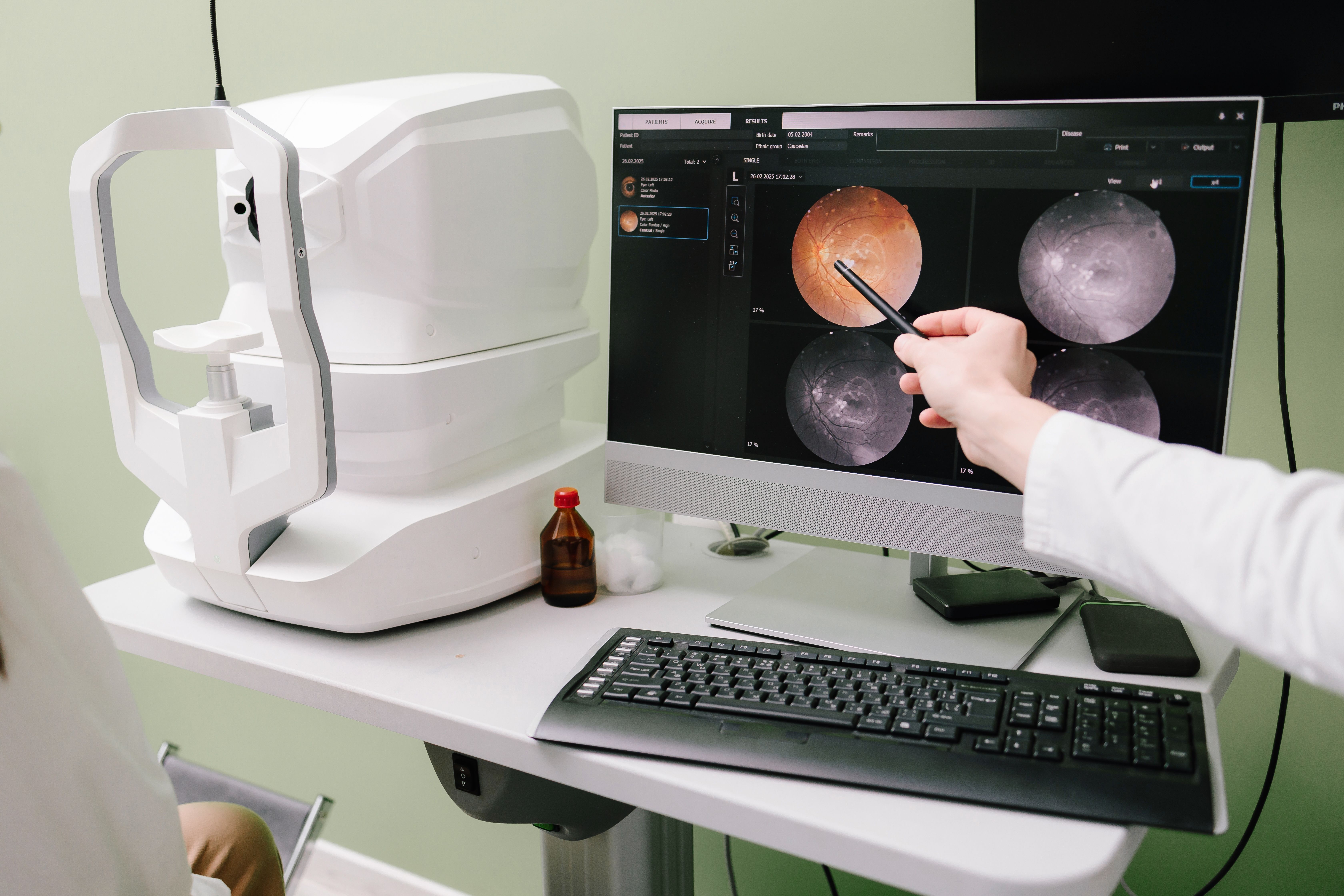
At Texas State Optical, advanced diagnostic tools such as Optical Coherence Tomography (OCT) and Optos widefield imaging play a critical role in protecting your long-term vision.
Why Early Detection Matters
Glaucoma and macular degeneration are leading causes of permanent vision loss, but early intervention can slow or even prevent progression. Traditional eye exams are essential, but retinal imaging provides a deeper, more detailed view of the structures inside the eye - revealing changes that are not always visible during a standard exam.
By capturing high-resolution images of the retina and optic nerve, your eye doctor can detect subtle abnormalities long before symptoms arise.
How Retinal Imaging Helps Detect Glaucoma
Glaucoma damages the optic nerve, often due to increased eye pressure, and typically progresses without pain or early vision changes. Retinal imaging is especially valuable because it allows doctors to monitor the optic nerve and surrounding nerve fiber layers with exceptional precision.
Optical Coherence Tomography (OCT) and Glaucoma
OCT uses light waves to create detailed cross-sectional images of the retina and optic nerve. This technology measures the thickness of the retinal nerve fiber layer, which is one of the earliest structures affected by glaucoma. Even minor thinning can be detected, enabling diagnosis before noticeable vision loss occurs.
OCT also helps track changes over time, allowing your optometrist to determine whether glaucoma is stable or progressing and to adjust treatment accordingly.
How Retinal Imaging Detects Macular Degeneration
Macular degeneration affects the macula, the part of the retina responsible for sharp, central vision needed for reading, driving, and recognizing faces. Early stages may not cause obvious symptoms, making imaging essential for early diagnosis.
OCT and Macular Health
OCT provides detailed images of the macula, helping detect early signs of macular degeneration such as fluid buildup, thinning, or structural changes. This level of detail allows eye doctors to distinguish between dry and wet forms of the disease and determine the most appropriate management plan.
Optos Imaging
Optos imaging captures an ultra-wide view of the retina - up to 200 degrees - in a single image. This allows doctors to see areas of the retina that traditional imaging may miss. For macular degeneration, Optos imaging helps identify peripheral retinal changes and overall retinal health, offering a more comprehensive picture of eye disease risk.
A Comfortable, Non-Invasive Experience
Both OCT and Optos imaging are fast, painless, and non-invasive. In many cases, dilation may not be required, making the process more comfortable and convenient for patients. The images are available immediately, allowing your optometrist to explain findings clearly and answer questions during your visit.
Start Protecting Your Vision Today
Retinal imaging has revolutionized the early detection and management of glaucoma and macular degeneration. By identifying subtle changes in the retina and optic nerve before symptoms appear, technologies like OCT and Optos imaging help preserve vision and support lifelong eye health. Regular comprehensive eye exams that include advanced imaging are one of the most effective ways to protect your sight.
Your vision is too important to leave to chance. Schedule a comprehensive eye exam with Texas State Optical today and ask about advanced retinal imaging. Visit our office in Richmond, Texas, or call (346) 210-5200 to book an appointment today.
Quick Links
Helpful Articles
article_category: cosmetic
article_category: eye surgery co-management
article_category: products
article_category: vision therapy
article_category: dogs
article_category: technology
article_category: general
article_category: eye health
article_category: aesthetics
article_category: services
article_category: faqs
article_category: exotic
article_category: large animal
article_category: contact lenses
article_category: surgical procedures
article_category: health
article_category: psychiatry
article_category: conditions
article_category: restorative
article_category: preventative
article_category: cats
article_category: eyeglasses
article_category: ocular disease management
[]
Flowbite is an open-source library of interactive components built on top of Tailwind CSS including buttons, dropdowns, modals, navbars, and more.
Check out this guide to learn how to get started and start developing websites even faster with components on top of Tailwind CSS.

All Eyecare Services
We offer a wide variety of eye care services to the Richmond community. Contact us with any questions about our services.

Keep In Touch
For non-urgent questions or to learn more about our services, contact us today!
Contact Information
Office Hours
- Monday 9:00 AM - 6:00 PM
- Tuesday 9:00 AM - 7:00 PM
- Wednesday 9:00 AM - 6:00 PM
- Thursday 9:00 AM - 7:00 PM
- Friday 9:00 AM - 6:00 PM
- Saturday 8:30 AM - 4:00 PM
- Sunday Closed
- Monday 9:00am - 6:00pm
- Tuesday 9:00am - 6:00pm
- Wednesday 9:00am - 6:00pm
- Thursday 9:00am - 6:00pm
- Friday 9:00am - 6:00pm
- Saturday 10:00am - 2:00pm
- Sunday Closed
- Monday 8:00am - 5:00pm
- Tuesday
- Wednesday
- Thursday
- Friday
- Saturday
- Sunday
© 2026 Texas State Optical. All rights Reserved - Accessibility Statement - Privacy Policy - Sitemap
Managed and Designed by ![]()